Secondary Structure Diagram From Pdb Four Levels Of Protein
How to "linearize" or "unfold" a pdb format structure to secondary Secondary structure predicted by the pdbsum database. helices labelled Sequence pdb entities secondary rcsb structureid
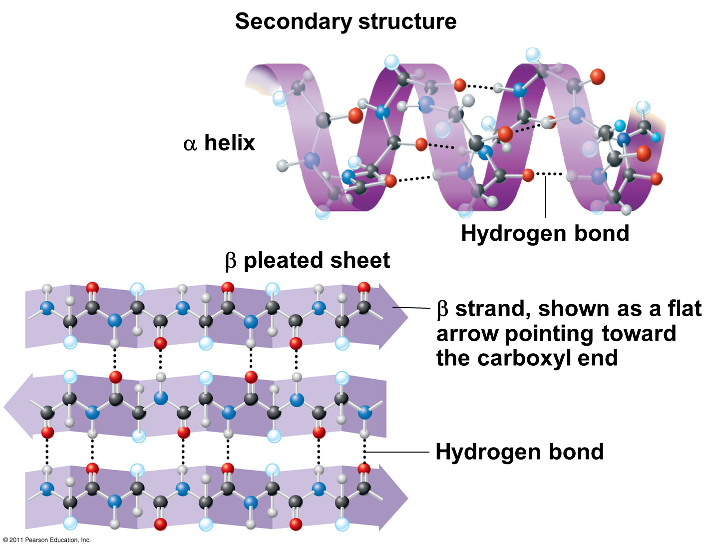
Protein secondary structure showing α -helix | Download Scientific Diagram
Sequence display for secondary structure entities in pdb model 3guu Protein primary structure diagram Pdb plot secondary sequence chain structure do like draw ribbon 0k strucure views
Proteins struktur helix sheet microbiology macromolecules chemistry hydrogen major bonds chain labeled kompas tertiary acids biochemistry spiral chapter sekunder libretexts
Protein secondary bonds interactions hydrogen covalent noncovalentPdb-101: learn: guide to understanding pdb data: computed structure models Protein structure & function (1.3.3)Secondary structure model.
Secondary structure predictions. predicted secondary structures for theLevels of structures of proteins (a) secondary structure predicted based on the templates (pdb id: 1tgsCh103 – chapter 8: the major macromolecules – chemistry.

How can i make a pdb file from a predicted secondary structure for a
Secondary structure protein proteins polypeptide amino acids bonds hydrogen pleated bond structures peptide biology sheets many together quaternary example levelsSecondary structure alignment and structural comparisons. a, the Solved the diagram of the secondary structure shown here isThe schematic representation of secondary structure prediction and.
How do you plot secondary structure like pdb?Sequence display for secondary structure entities in pdb model 3zpx Protein secondary structure prediction serviceStructure secondary protein prediction service proteomics creative.

[solved] how many hydrogen bonds involving the backbone co and nh can
The pdb annotated by secondary domains that contain multiple dataAssigning secondary structural elements from pdb files using dssp Orders of protein structureGraphs depicts publication.
Secondary structure analysis. the graphs depicts the secondary| the schematic diagram of the secondary structure information Secondary structure in pdb homologs for simple lcrs by type. for eachSecondary structure.

Pdb predicted step1 step2
Secondary structures of protein pdb id: 6bi6.Secondary structure The biologs: cape 1: proteinsProtein secondary structure showing α -helix.
Pdb dihedral backbone linearize unfoldFour levels of protein structure and examples Protein secondary structure predictionAnalysis of secondary structure. secondary structure was analyzed using.

Protein design
Alignment structure structural comparisonsComputing the secondary structure from pdb entries for each time frame .
.

![[Solved] How many hydrogen bonds involving the backbone CO and NH can](https://i2.wp.com/cdn.testbook.com/images/production/quesImages/qImage646f23abed4806260c7a04d2.png)
[Solved] How many hydrogen bonds involving the backbone CO and NH can

Sequence display for secondary structure entities in PDB model 3GUU

Secondary Structure

Secondary structure alignment and structural comparisons. A, the

Four levels of protein structure and examples - Biology Brain
The BioLogs: CAPE 1: Proteins

Secondary structures of protein PDB ID: 6BI6. | Download Scientific Diagram